Figure 6
Delayed Diagnosis of Early-onset Sarcoidosis: A Case Report and Literature Review
Mutibah Ali Al-essi*, Lujain Salah Binkhamis, Samah Mohammed Aljohani and Nora Mohammad Alzahrani
Published: 18 January, 2024 | Volume 7 - Issue 1 | Pages: 001-005
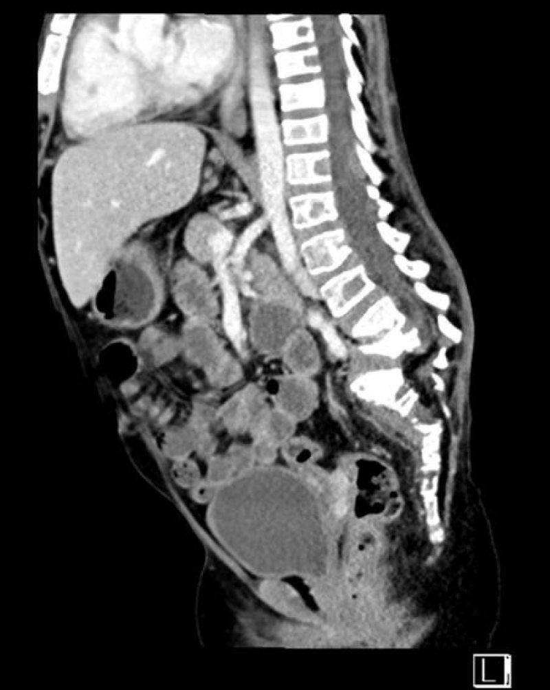
Figure 6:
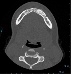
Figure 6: Abdominal and pelvic computed tomography without contrast revealed diffuse abnormal lytic and sclerotic bone lesions involving the entire visualized part of the spine, including the lower dorsal spine. Notably, it depicted a compression fracture or gibbus deformity of the lower vertebral bodies L4 and L5, along with malalignment and displacement of the lumbosacral spine.
Read Full Article HTML DOI: 10.29328/journal.japch.1001061 Cite this Article Read Full Article PDF
More Images
Similar Articles
-
Delayed Diagnosis of Early-onset Sarcoidosis: A Case Report and Literature ReviewMutibah Ali Al-essi*, Lujain Salah Binkhamis, Samah Mohammed Aljohani, Nora Mohammad Alzahrani. Delayed Diagnosis of Early-onset Sarcoidosis: A Case Report and Literature Review. . 2024 doi: 10.29328/journal.japch.1001061; 7: 001-005
-
Case Report: An Elusive Case of Septic ArthritisGeorgina George Balyorugulu*, Shabani Yusuph, Rahma Majaliwa, Mpuya Innocent, Fikiri Martine, Fatma Said, Rogatus Kabyemera, Patrick Ngoya, Jeremiah Seni. Case Report: An Elusive Case of Septic Arthritis. . 2024 doi: 10.29328/journal.japch.1001067; 7: 045-051
Recently Viewed
-
Do Fishes Hallucinate Human Folks?Dinesh R*,Sherry Abraham,Kathiresan K,Susitharan V,Jeyapavithran C,Paul Nathaniel T,Siva Ganesh P. Do Fishes Hallucinate Human Folks?. Arch Food Nutr Sci. 2017: doi: 10.29328/journal.afns.1001003; 1: 020-023
-
Assessment of Redox Patterns at the Transcriptional and Systemic Levels in Newly Diagnosed Acute LeukemiaAna Carolina Agüero Aguilera, María Eugenia Mónaco, Sandra Lazarte, Emilse Ledesma Achem, Natalia Sofía Álvarez Asensio, Magdalena María Terán, Blanca Alicia Issé, Marcela Medina, Cecilia Haro*. Assessment of Redox Patterns at the Transcriptional and Systemic Levels in Newly Diagnosed Acute Leukemia. J Hematol Clin Res. 2024: doi: 10.29328/journal.jhcr.1001029; 8: 017-023
-
Assessment of Indigenous Knowledge on Using of Traditional Medicinal Plants to Cure Human Diseases in South Omo Zone Baka Dawla Ari District, Kure and Bitsmal South EthiopiaGizaw Bejigo*. Assessment of Indigenous Knowledge on Using of Traditional Medicinal Plants to Cure Human Diseases in South Omo Zone Baka Dawla Ari District, Kure and Bitsmal South Ethiopia. J Plant Sci Phytopathol. 2024: doi: 10.29328/journal.jpsp.1001132; 8: 048-054
-
Nanoencapsulated Extracts from Leaves of Bauhinia forficata Link: In vitro Antioxidant, Toxicogenetic, and Hypoglycemic Activity Effects in Streptozotocin-induced Diabetic MiceBárbara Verônica Cardoso de Souza, Alessandra Braga Ribeiro*, Rita de Cássia Meneses Oliveira, Julianne Viana Freire Portela, Ana Amélia de Carvalho Melo Cavalcante, Esmeralda Maria Lustosa Barros, Luís Felipe Lima Matos, Tarsia Giabardo Alves, Maria. Nanoencapsulated Extracts from Leaves of Bauhinia forficata Link: In vitro Antioxidant, Toxicogenetic, and Hypoglycemic Activity Effects in Streptozotocin-induced Diabetic Mice. Arch Pharm Pharma Sci. 2024: doi: 10.29328/journal.apps.1001063; 8: 100-115
-
GELS as Pharmaceutical Form in Hospital Galenic Practice: Chemico-physical and Pharmaceutical AspectsLuisetto M*,Edbey Kaled,Mashori GR,Ferraiuolo A,Fiazza C,Cabianca L,Latyschev OY. GELS as Pharmaceutical Form in Hospital Galenic Practice: Chemico-physical and Pharmaceutical Aspects. Arch Surg Clin Res. 2025: doi: 10.29328/journal.ascr.1001084; 9: 001-007
Most Viewed
-
Evaluation of Biostimulants Based on Recovered Protein Hydrolysates from Animal By-products as Plant Growth EnhancersH Pérez-Aguilar*, M Lacruz-Asaro, F Arán-Ais. Evaluation of Biostimulants Based on Recovered Protein Hydrolysates from Animal By-products as Plant Growth Enhancers. J Plant Sci Phytopathol. 2023 doi: 10.29328/journal.jpsp.1001104; 7: 042-047
-
Sinonasal Myxoma Extending into the Orbit in a 4-Year Old: A Case PresentationJulian A Purrinos*, Ramzi Younis. Sinonasal Myxoma Extending into the Orbit in a 4-Year Old: A Case Presentation. Arch Case Rep. 2024 doi: 10.29328/journal.acr.1001099; 8: 075-077
-
Feasibility study of magnetic sensing for detecting single-neuron action potentialsDenis Tonini,Kai Wu,Renata Saha,Jian-Ping Wang*. Feasibility study of magnetic sensing for detecting single-neuron action potentials. Ann Biomed Sci Eng. 2022 doi: 10.29328/journal.abse.1001018; 6: 019-029
-
Pediatric Dysgerminoma: Unveiling a Rare Ovarian TumorFaten Limaiem*, Khalil Saffar, Ahmed Halouani. Pediatric Dysgerminoma: Unveiling a Rare Ovarian Tumor. Arch Case Rep. 2024 doi: 10.29328/journal.acr.1001087; 8: 010-013
-
Physical activity can change the physiological and psychological circumstances during COVID-19 pandemic: A narrative reviewKhashayar Maroufi*. Physical activity can change the physiological and psychological circumstances during COVID-19 pandemic: A narrative review. J Sports Med Ther. 2021 doi: 10.29328/journal.jsmt.1001051; 6: 001-007

HSPI: We're glad you're here. Please click "create a new Query" if you are a new visitor to our website and need further information from us.
If you are already a member of our network and need to keep track of any developments regarding a question you have already submitted, click "take me to my Query."


